

Establishing the definitive visual gold standard for neurosurgical pathology.

the story
Engaged by the Congress of Neurological Surgeons (CNS), we architected a specialized series of 14 foundational didactic illustrations for the CNS Nexus platform. Designed to serve as the "gold standard" representations of complex neurosurgical conditions, these works provide a definitive visual anchor for a case-based learning exchange utilized by the global neurosurgical community. Spanning trauma, oncology, pediatrics, and epilepsy, the series translates highly variable clinical data into a clear, standardized educational baseline.
timeframe
2019 – 2021
tools
Illustrator • Photoshop • Procreate
services
Medical Illustration • Didactic Design • Educational Standardization
problem
Neurosurgical education relies heavily on raw radiological scans, which are inherently variable and difficult to standardize for broad didactic application. To bridge this gap, the CNS Nexus platform required a comprehensive library of archetypal illustrations, enabling both residents and attending surgeons to rapidly and accurately recognize the structural hallmarks of specific traumas, tumors, and congenital anomalies.
solution
Collaborating closely with subject matter experts across multiple institutions, we engineered a unified suite of high-fidelity pathological renderings. By isolating and standardizing specific clinical features—such as hematoma progression, skull base fracture levels, and neuromodulation pathways—this system establishes a definitive visual baseline that perfectly complements raw imaging data, removing visual noise to reveal pure structural truth.
Pathology Explorer: The CNS Nexus Library
An interactive review of the didactic "gold standard" pathological illustrations created for the Congress of Neurological Surgeons (CNS).
Select a category below to explore the standardized presentations of trauma, pediatric anomalies, epilepsy, and neuro-oncology.
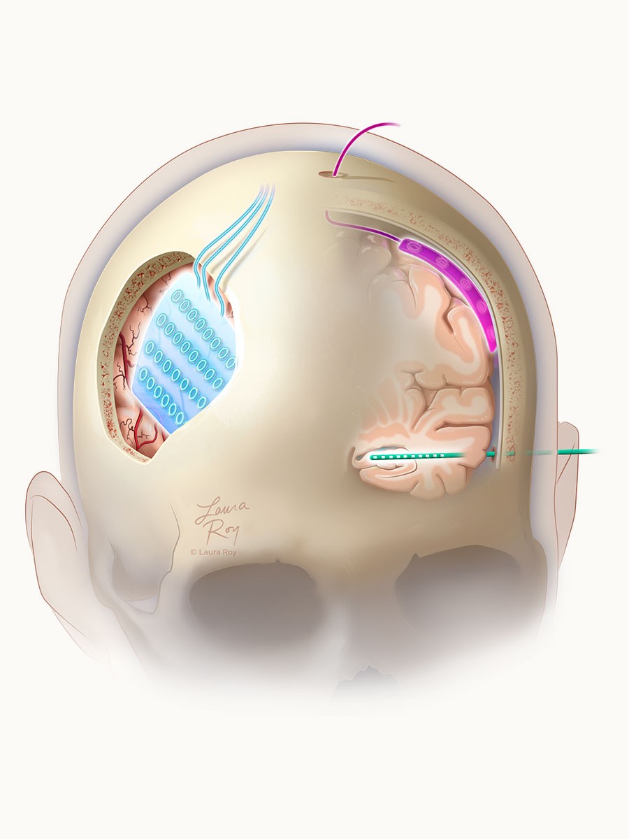
Monitoring
Invasive intracranial monitoring utilizes depth electrodes or subdural grids to precisely map epileptogenic zones prior to surgical resection.
— Maryam Rahman, MD, MS